Intestine-on-a-chip for Drug Metabolism and Infectious Disease Modeling
Innovative Products and Technologies
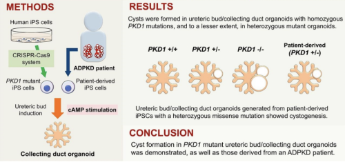
Microfluidic organ-on-a-chip technique has recently gained attention for modeling various organs, including the small intestine. A variety of intestine chip designs have been implemented with the double-layer channel separated by a porous PDMS membrane as the most popular option. Available on the market devices are usually composed of epithelial cells in the top channel, endothelial cells at the bottom, and mediums flowing through the two channels to recreate shear stress the intestinal cells experience postnatally. The fluid flowing through the bottom channel is normally designed to emulate the blood. However, the researchers at Kyoto University suspected that mimicking interstitial flow, essential for embryonic organogenesis, in the bottom channel instead could be the key to producing a physiologically relevant intestine-on-a-chip model. They tested this hypothesis and, using human pluripotent stem cells (PSCs), successfully developed a micro-small intestine system that faithfully recapitulates small intestine physiology.
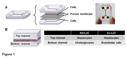
First, the researchers fabricated the microfluidic devicefrom polydimethylsiloxane (PDMS) using a soft lithographic method. The deviceconsisted of two layers of microchannels separated by a semipermeable membrane with 3.0-μm pores. The researchers then seeded humanPSCs,embryonic stem (ES) or induced pluripotent stem (iPS) cell lines, on cell culture plates and treated them with an intestinal differentiation medium. The cells were then injected into the top channel of the PDMS device for continued differentiation. To reproduce interstitial flow in the microfluidic device, the researchers performed experiments with various flow velocities and duration in the bottom channel of the device and optimized the medium flow to 30 μL/h from days 5 to 24 (Fig.1, Deguchi et al. 2024)
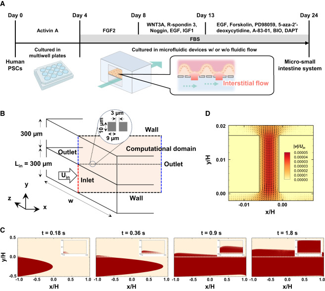
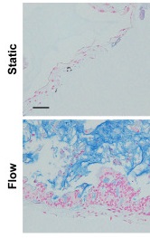
Figure 1. Differentiation procedure to generate micro-small intestine using human PSCs and the microfluidic device.Micro-small intestine systems were cultured under static or flow conditions.
➤Interstitial flow is recapitulated: multilayered structure is formed
| The researchers then confirmed that intestinal epithelial and mesenchymal cells differentiated simultaneously from the seeded PSCs in the micro-small intestine system indicating the recapitulation of the cell type diversity found in naturally formed tissues. Through histological and RNA/protein-based analyses, the researchers found that the interstitial flow generated in the device prompted primitive villi-like 3D structures made of epithelial cells to develop with the mesenchymal layer aligned under them. Moreover, this isthe first intestine-on-a-chip system which enabled to reconstruct a multilayered structure consisting of a mucus layer, intestinal epithelial layer, mesenchymal cell layer, and ECM(Fig.2, Deguchi et al. 2024). |
Figure 2.Alcian blue staining reveals a mucus layer formed under flow conditions. |
➤ Intestine-on-a-chip as a platform for drug metabolism and disease modeling
Next, the researchers showed that the interstitial flow increased the protein levels of drug-metabolizing enzymes. They also showed that ANPEP, a receptor of the HCoV-229E human coronavirus, known to cause gastrointestinal symptoms, was expressed at a higher level and correctly localized to the apical side of the intestinal epithelium. A detailed examination of infected tissues by RNA sequencing confirmed that the system accurately reproduces the viral infection in the small intestine. Therefore, the researchers confirmed that their micro-small intestine system is useful for pharmaceutical and infectious disease research.
Intellectual property status
Granted Patent
Patent number :
Where : Japan
Current development status
Laboratory prototypes
Desired business relationship
Patent licensing
Technology development
Kyoto University was founded in 1897, the second university to be established in Japan. Kyoto University is among 10 National Designated Universities in Japan. It boasts 18 graduate schools, 10 faculties, 12 research institutes, and 26 centers and other establishments. Research conducted at Kyoto University spans the full spectrum of fields from social to natural science.
The outstanding research conducted at Kyoto University gives birth to useful technologies that could greatly benefit society. IAC (Office of Institutional Advancement and Communications) was established at Kyoto University to bridge the gap between researchers and industry. We facilitate joint research, technology transfer, creation of university startups, and provide entrepreneurial education. We are building a strong network of global industry partners to make sure basic research reaches the market.
Create your free account to connect with Larisa Sheloukhova and thousands of other innovative organizations and professionals worldwide
Send a request for information
to Larisa
Technology Offers on Innoget are directly posted
and managed by its members as well as evaluation of requests for information. Innoget is the trusted open innovation and science network aimed at directly connect industry needs with professionals online.
Need help requesting additional information or have questions regarding this Technology Offer?
Contact Innoget support